Cells and Microscopes
Animal Cells:
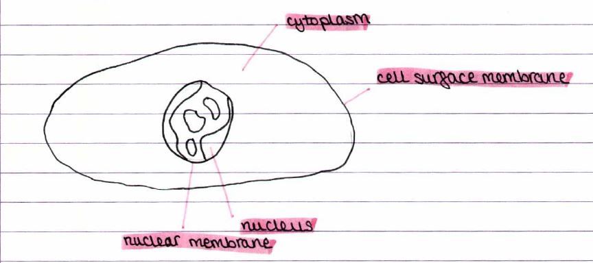
Nucleus: Control centre of the cell. Contains DNA, the genetic information.
Nuclear membrane: Controls what enters and leaves the nucleus.
Cell surface membrane: Controls what enters and leaves the cell.
Cytoplasm: A jelly-like part of the cell where metabolic reactions happen.
Plant Cells:
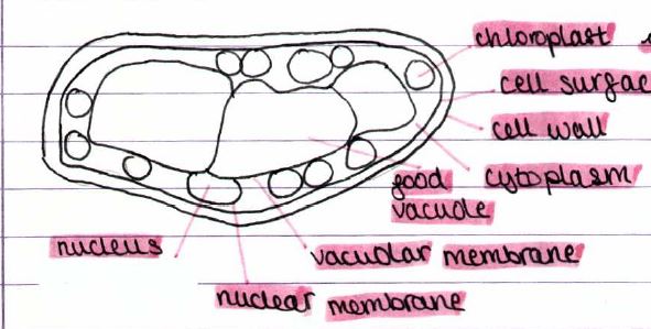
- Chloroplast: The site of photosynthesis.
- Cell wall: Made of cellulose. It's strong and inelastic, stopping the cell from taking in too much water and bursting.
- Food vacuole: Holds a fluid known as cell sap, which stores useful waste molecules, primarily water.
- Vacuolar membrane: Regulates what enters and exits the vacuole.
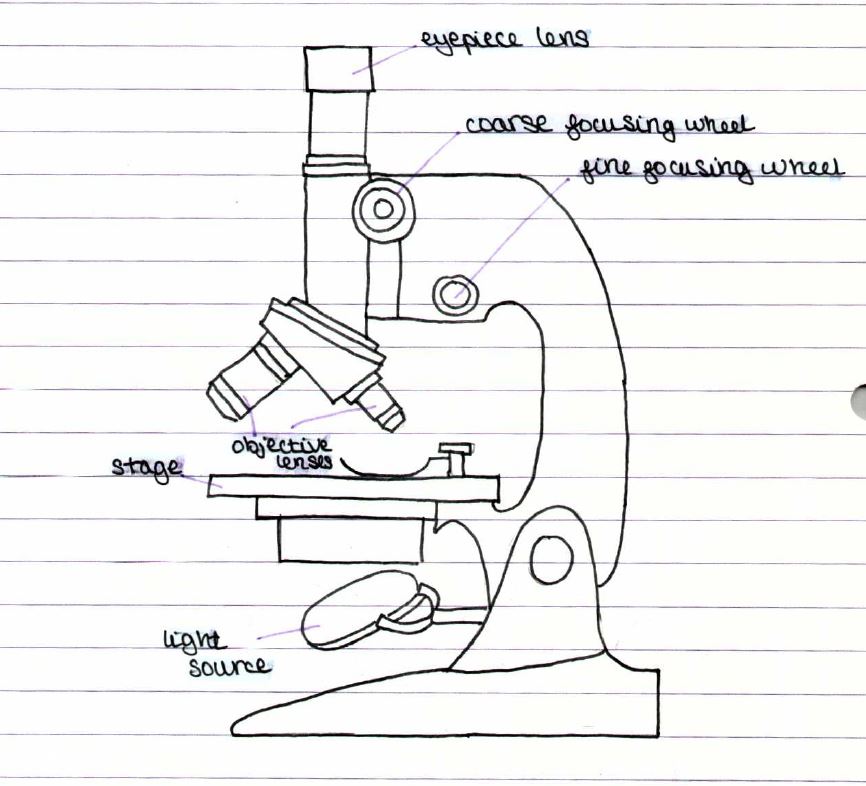
Light Microscopes
B1.1a
- Can be used to magnify an image up to x1500, making it larger than the actual cell size.
- They use two different lenses to enlarge the image of the specimen under observation.
- Overall magnification: Eyepiece lens x Objective lens
Diagram of a Microscope:
Calculations
in microscopy
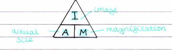
Image size: Actual size x Magnification
Magnification: Image size ÷ Actual size
Actual size: Image size ÷ Magnification
Examples:
1) If actual size = 18 mm and image size = 55 mm
Magnification: 55 ÷ 18 = 3.05 mm.
2) Magnification = x1000 and image size = 4.2 mm
Actual size: 4.2 ÷ 1000 = 0.0042 mm
Micrometres and Nanometres
- A micrometre, μm, is the size of a mm divided into 1000 equal divisions.
- It's 10⁻⁶ of a metre.
- A typical animal cell is 40 mm wide; this varies considerably.
- A nanometre, nm, is the size of a μm divided into 1000 equal parts.
- It's 10⁻⁹ of a metre.
- Cell membranes are 10 nm wide. Viruses are about 40 to 100 nm in diameter.
Examples
of Micrometre Questions
1) a. 39 mm = 39,000 μm
b. Magnification = x900
c. Actual size = 39000 ÷ 900 = 43 μm.
2) a. Scale bar 27 mm = 6 μm (actual size) → 27,000 μm
b. 27,000 ÷ 6 = 4,500 → magnification = x4500
c. Actual size = 46,000
a = i ÷ m
46,000 ÷ 4,500 = 10.2 μm → actual size
Check the answer with the scale bar to make sure it's rational.
3) a. Scale bar = 50 μm (actual size) Image size: 20 mm
magnification: 
b. image length = 26 mm = 26,000
a = i ÷ m
26,000 ÷ 400 = 65 μm
The actual size of the cell is 65 micrometres.
Using Microscopes
Making Slides
1) Place the specimen onto the glass slide, ensuring it is as flat and smooth as possible.
2) Add one drop of iodine.
3) Put the coverslip on top. Carefully lower this down and press gently. Use filter paper to mop up any excess stain that spills out to the sides.
4) Look at the slide using a light microscope. Always start with the lowest power objective lens and position the stage as close to the objective lens as possible. Use focusing wheels to then get the image focused.
Resolution:
- Light microscopes have a maximum magnification and resolution.
- Magnification: This refers to how many times larger the image is.
- Resolution: The ability to distinguish between two points.
B1.1c Electron Microscopes:
- They produce images with a higher magnification and resolution than light microscopes.
- A beam of electrons passes through the specimen instead of light.
- Instead of using lenses, electron microscopes use magnets.
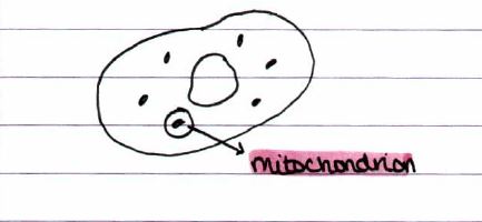
- They allow us to see more detail (organelles) within cells. E.g., mitochondria
Advantages: Higher magnification and resolution, allowing us to see different organelles.
Disadvantages: The device is highly costly, requires extensive training for operation, necessitates a dead specimen in a complete vacuum, and produces only black and white images.
TEM: The Transmission Electron Microscope has a magnification of up to x 1,000,000. Allows us to see inside cells.
SEM: The Scanning Electron Microscope is a tool used to examine the surface of cells.
Parts of cells
b1.1b
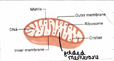
Mitochondria:
- Organelles found in plant and animal cells.
- Around 5 μm.
- Creates energy from aerobic respiration.
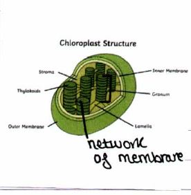
Chloroplasts:
- Found in plant cells.
- Around 10 μm.
- It captures sunlight and serves as the location for photosynthesis.
Ribosomes:
- Plant, animal, and bacterial cells contain this substance.
- They consist of two subunits, each of which builds proteins from amino acids.
- Protein synthesis varies from 20 to 30 nm.
- Metabolic reactions all take place in the cytoplasm apart from aerobic respiration in the mitochondria.
Cell Membranes:
- This system controls what can enter and leave the cell.
- The cell houses numerous molecules that function as receptors. They detect signals from the cell's environment and send messages to the cell.
Prokaryotes
- Prokaryotes: bacteria are significantly smaller than eukaryotes: plant and animal cells.
- The main structural difference between the two of them is that prokaryotes don't have a nucleus.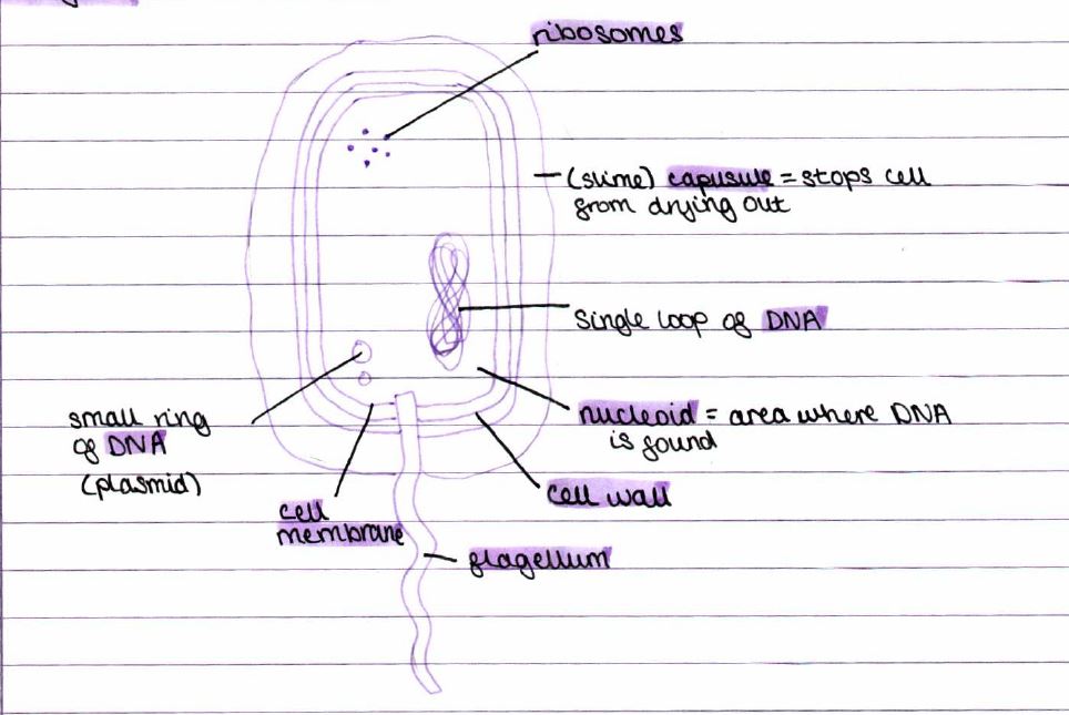
- Prokaryotes don't have a nucleus, but eukaryotes do.
- Prokaryotes don't have membrane-bound organelles, but eukaryotes do.
- They both have genetic material (DNA) and have a cell surface membrane and cytoplasm.
- Prokaryotes can only be single-celled, whereas eukaryotes can be single- and multicelled.
Order of Magnitude:
metre = 1 x 10⁰
centimetre = 1 x 10⁻²
millimetre = 1 x 10⁻³
micrometre = 1 x 10⁻⁶.
nanometre = 1 x 10⁻⁹